Citation: Huang D, Chen E, Rupenthal ID, “Using Physical Forces to Enhance Ocular Drug Delivery”. ONdrugDelivery Magazine, Issue 82 (Jan 2018), pp 6-9.
Di Huang, Erica Chen and Ilva Rupenthal discuss the possibilities offered by physical force-based methods of enhancing the efficacy of ophthalmic drug delivery by providing a way past the eye’s natural barriers.
This piece reviews a 2017 article the authors published in Advanced Drug Delivery Reviews.1
INTRODUCTION
Delivering therapeutics to specific sites in the eye whilst also achieving effective drug concentrations is a difficult task, due to a number of inherent anatomical and physiological ocular barriers, including the cornea, the sclera, Bruch’s-choroid complex and the blood-retinal barrier. These barriers not only protect the eye from invasion by foreign substances, but also regulate the intraocular milieu, which is essential for the eye’s physiological function. However, these barriers also pose a major obstacle to efficient drug delivery, overcoming which remains a major challenge to improve ocular drug bioavailability, with various strategies having been investigated over recent decades.
Formulation-based approaches include chemical penetration enhancers, prodrugs and drug delivery carriers such as liposomes and nano- or microparticles able to penetrate the ocular barriers. Physical force-based methods, initially utilised in transdermal drug delivery, generally require a power-driven physical device to deliver energy to the barriers, thereby enhancing transient drug transport. Compared with formulation-based approaches, physical force-based strategies may allow greater control over the drug dosage whilst also offering the possibility to record parameter information. Here follows a brief overview of physical methods (Figure 1), including iontophoresis, sonophoresis and microneedles, which can enhance drug penetration by transiently disrupting the ocular barriers in a minimally or non-invasive manner.1

Figure 1: Overview of physical force-based methods for ocular drug delivery enhancement.
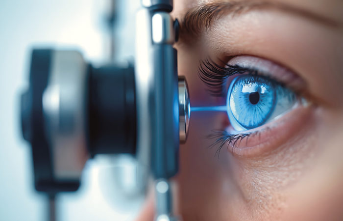
IONTOPHORESIS
Iontophoresis, application of a low-intensity electrical current, enhances drug delivery across biological membranes by causing electrorepulsion and electro-osmosis of the drug molecule.2,3 Electrorepulsion primarily applies to the movement of ionic drugs,4 while electro-osmosis can enhance the transport of both neutral and charged molecules by convective solvent flow.5 The relative contribution of electrorepulsion and electro-osmosis depends on both the physicochemical characteristics of the drug (e.g. size, charge and charge to molecular-weight ratio) and the electrical properties of the biological membrane. Iontophoretic permeability enhancement of small, charged molecules is mainly governed by electrorepulsion, along with a minor contribution by electro-osmosis, whereas for macromolecules the mechanism is highly dependent on the charge to molecular-weight ratio.
“Compared with formulation-based approaches, physical force-based strategies may allow greater control over the drug dosage whilst also offering the possibility to record parameter information…”
The basic design of ocular iontophoretic devices consists of a power source and two electrodes: the donor electrode (an ocular applicator or eye cup) and the return electrode. The drug is filled into the applicator and the return electrode is placed on the forehead to form an electrical circuit. The EyeGate® II Delivery System (EyeGate Pharma, MA, US), an annular shaped silicone probe with a 0.5 cm2 contact area and a 13 mm inner diameter used for transscleral iontophoresis, for example, is currently being investigated in a number of clinical trials for delivery of a sustained release dexamethasone formulation (EGP-437) in the treatment of anterior uveitis, dry eye and macular oedema, as well as for prevention of ocular inflammation in patients who have undergone cataract surgery.
SONOPHORESIS
Sonophoresis, also called ultrasound, involves the application of a sound field at frequencies higher than 20 kHz to improve drug transport across biological membranes, including ocular barriers.6 It has been utilised in the field of ophthalmology for decades but primarily as a diagnostic imaging tool.7 However, therapeutic ultrasound has recently emerged as an option to treat glaucoma by cyclocoagulation8 or to enhance ocular drug uptake. The mechanisms for ultrasound enhanced drug delivery take into account non-thermal (e.g. cavitation, acoustic streaming and mechanical stress) and thermal effects with ultrasound parameters, co-administration of microbubbles and drug characteristics, all having an effect on delivery efficacy. Cavitation is generally considered the predominant factor for enhanced drug delivery and is defined as the formation of microbubbles due to an acoustic pressure gradient within the coupling medium. Continuous pulsation of cavitation microbubbles over many pressure cycles without any collapse is considered stable cavitation, while the growth and collapse of microbubbles within a few pressure cycles is associated with inertial cavitation.
“The basic design of ocular iontophoretic devices consists of a power source and two electrodes: the donor electrode and the return electrode. The drug is filled into the applicator and the return electrode is placed on the forehead to form an electrical circuit…”
Corneal permeability enhancement is generally a result of stable cavitation at low ultrasound intensities, whereas both stable and inertial cavitation play important roles at higher ultrasound strengths.6 Transsclerally applied ultrasound results in the formation of transport channels and the modification of the proteoglycan fibre morphology, without significantly disturbing the collagen network. Here, low intensity ultrasound produces sufficient acoustic pressure to generate stable oscillating microbubbles, resulting in microstreaming with higher intensities being required to achieve inertial cavitation.9 Due to the sensitivity of the ocular tissues, it is incredibly important not to sacrifice patient safety, even if doing so could potentially achieve greater treatment efficacy. The eye’s temperature increase due to absorption of sound waves is of particular concern; the thermal safety requirements for diagnostic ultrasound allow a maximum temperature increase of 1.5 °C.
The majority of studies published to date have investigated transcorneal ultrasound in combination with microbubbles; however, a limited number of studies also exist on the application of ultrasound for enhanced drug delivery to the posterior segment of the eye. Recently, transscleral ultrasound has also been combined with the use of nanocarriers, in order to enhance the vitreous diffusion and retinal permeability of peptide loaded nanoparticles following intravitreal injection, thus achieving higher retinal drug concentrations.10 However, so far, there has been no evidence that ultrasound can enhance retinal permeability upon extraocular administration in large animal models or humans.
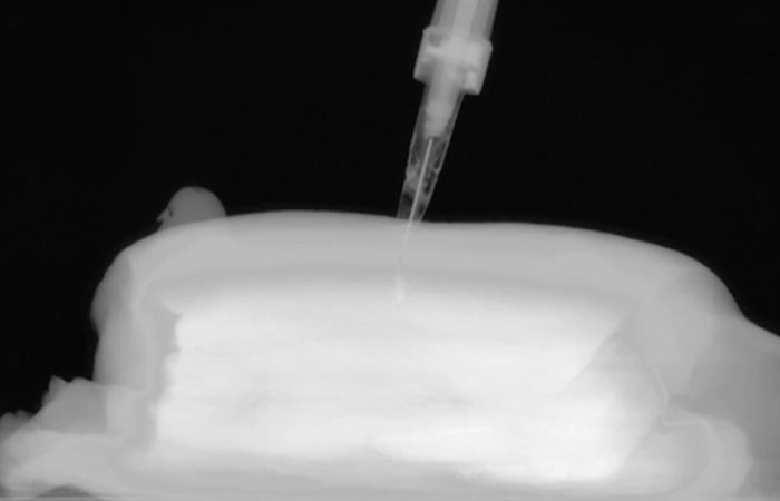
MICRONEEDLES
Microneedles (MNs) are micrometre sized needles, or arrays of such, fabricated by adapting microelectronics tools. Applying MNs to biological membranes can create tiny transport pathways, thereby allowing drugs to permeate across these barriers. To date, numerous MN fabrication approaches have been utilised, resulting in a variety of shapes, sizes, materials and configurations.11
According to their delivery mechanism, ocular MNs can be categorised into four types:
- Solid MNs, able to create micropores in the ocular surface through which therapeutics can diffuse after MN removal.
- Drug-coated MNs, with the drug coating dissolving and diffusing into the eye after insertion.
- Dissolving MNs, which disintegrate over time, thus releasing the matrix encapsulated drug into the ocular tissues.
- Hollow MNs, able to infuse pressure-driven liquid drug formulations.
Enhanced drug delivery into the cornea and anterior segment of the eye can be achieved by insertion of MNs across the corneal epithelium, the main barrier to penetration encountered after topical eye drop administration, to deposit drugs directly into the corneal stroma. Solid stainless steel MNs, coated with different compounds ranging from small drugs to macromolecules, were inserted into rabbit corneas in vivo providing up to 60-fold higher bioavailability compared with the topically administered drugs.12 However, in vivo application is still challenging due to the corneal curvature and lack of a supporting pressure during MN insertion.
“Therapeutic ultrasound has recently emerged as an option to treat glaucoma by cyclocoagulation or to enhance ocular drug uptake…”
Various polymeric MNs have found great use in intrascleral drug delivery. Unlike solid or hollow MNs, dissolving MNs minimise accidental retinal damage, whilst also remaining as a depot for sustained drug delivery. Thakur, et al,13 developed a simple and cost-effective mouldcasting method for fabrication of rapidly dissolving MNs, using polyvinylpyrrolidone, which efficiently penetrated the outer scleral layers, thus enhancing intrascleral permeation of macromolecules.
Currently the most advanced ocular MN application includes drug delivery into the suprachoroidal space, a potential space between sclera and choroid. Drug solutions injected into this space can flow circumferentially around the eye, with suspensions of particles up to 1 μm having been successfully delivered into the suprachoroidal space of rabbit, pig and human eyes.14 Clearside Biomedical (GA, US) initially evaluated the efficacy of suprachoroidal delivery using hollow MNs for the treatment of acute posterior segment uveitis by injecting triamcinolone acetonide into the suprachoroidal space of living pigs.15 The company subsequently completed a Phase II clinical trial to determine the safety and efficacy of suprachoroidally administered, proprietary, non-preserved triamcinolone acetonide (Zuprata™) via 1000 μm long MNs in subjects with macular oedema associated with non-infectious uveitis. Currently participants are being recruited for a Phase III study for further efficacy evaluation. Clearside Biomedical also completed a Phase II clinical trial in subjects with macular oedema following retinal vein occlusion and is currently investigating suprachoroidal application of a small tyrosine kinase inhibitor (Axitinib™) in the treatment of wet age-related macular degeneration.
CONCLUSION
Safe and effective treatment of ocular diseases is a challenging task due to the presence of various protective ocular barriers and elimination mechanisms. A vast number of novel strategies have been utilised to overcome these barriers and improve drug delivery to the target site, thereby enhancing drug bioavailability and avoiding potential side effects. The enhancement of drug permeability, ease of application and minimally or non-invasive delivery characteristics render physical force-based methods an exciting option for the treatment of both anterior and posterior segment disorders. Iontophoresis is the most extensively investigated approach among these physical techniques so far, with numerous therapeutic agents (such as low molecular weight drugs, macromolecules and nanocarriers) already having been successfully delivered to various ocular tissues. Although the number of studies on ultrasound and MN mediated ocular drug delivery is still limited, both have specific advantages, including site-specific drug delivery to the ocular tissues as well as the possibility to combine them with sustained release particles. Further in vivo studies are required to understand the contribution of dominant mechanisms, optimise device design and parameter settings, and evaluate the feasibility and safety of repeated and long-term application of such methods in the clinical setting.
REFERENCES
- Huang D, Chen YS, Rupenthal ID, “Overcoming ocular drug delivery barriers through the use of physical forces”. Adv Drug Deliv Rev, 2017. (DOI: 10.1016/j.addr.2017.09.008.) Copyright © 2017 Elsevier.
- Kalia YN, Naik A, Garrison J, Guy RH, “Iontophoretic drug delivery”. Adv Drug Deliv Rev, 2004, Vol 56(5), pp 619-658.
- Guy RH et al, “Iontophoresis: Electrorepulsion and electro-osmosis”. J Control Release, 2000, Vol 64(1-3), pp 129-132.
- Marro D et al, “Contributions of electromigration and electro-osmosis to iontophoretic drug delivery”. Pharm Res, 2001, Vol 18(12), pp 1701-1708.
- Kasting GB, “Theoretical-models for iontophoretic delivery”. Adv Drug Deliv Rev, 1992, Vol 9(2-3), pp 177-199.
- Zderic V, Clark JI, Vaezy S, “Drug delivery into the eye with the use of ultrasound”. J Ultras Med, 2004, Vol 23(10), pp 1349-1359.
- Mundt GH Jr, Hughes WF Jr, “Ultrasonics in ocular diagnosis”. Am J Ophthalmol, 1956, Vol 41(3), pp 488-498.
- Aptel F, Lafon C, “Treatment of glaucoma with high intensity focused ultrasound”. Int J Hyperther, 2015, Vol 31(3), pp 292-301.
- Suen WL et al “Examination of effects of low-frequency ultrasound on scleral permeability and collagen network”. Ultras Med Biol, 2016, Vol 42(11), pp 2650-2661.
- Huang D, Chen YS, Rupenthal ID, “Ultrasound-mediated nanoparticle delivery across ex vivo bovine retina after intravitreal injection”. Eur J Pharmaceutics and Biopharmaceutics, 2017, Vol 119, pp 125-136.
- Donnelly RF, Raj Singh TR, Woolfson AD, “Microneedle-based drug delivery systems: microfabrication, drug delivery, and safety”. Drug Deliv, 2010, Vol 17(4), pp 187-207.
- Jiang J et al “Coated microneedles for drug delivery to the eye”. Invest Ophthalmol Vis Sci, 2007, Vol 48(9), pp 4038-4043.
- Thakur RR et al “Rapidly dissolving polymeric microneedles for minimally invasive intraocular drug delivery”. Drug Deliv Transl Res, 2016, Vol 6(6), pp 800-815.
- Patel SR et al, “Suprachoroidal drug delivery to the back of the eye using hollow microneedles”. Pharm Res, 2011, Vol 28(1), pp 166-176.
- Gilger BC et al, “Treatment of acute posterior uveitis in a porcine model by injection of triamcinolone acetonide into the suprachoroidal space using microneedles”. Invest Ophthalmol Vis Sci, 2013, Vol 54(4), pp 2483-2492.