
To Issue 136
Citation: Zhou Y, “Dissecting the Delicate Delivery Process for Ocular Gene Therapy”. ONdrugDelivery, Issue 136 (Aug 2022), pp 57–59.
Yongdong Zhou considers the challenges of ocular gene therapy delivery approaches.
“Ocular gene therapy has the potential to treat diseases previously considered untreatable and restore hope to millions of patients suffering from ocular issues.”
Gene therapy has emerged as one of the most promising areas of medicine in recent years. Not a year goes by without multiple breakthroughs in the field, and nowhere is there so much potential as in ocular gene therapy.
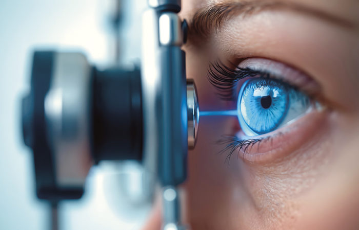
One of the major challenges of gene therapy has always been its delivery. It is incredibly difficult to insert genetic code into the correct cells, and consensus over the best means of delivery has evolved in recent years. This is particularly true for the eyes, where delivery is immensely complex.
The human eye is particularly suited to gene therapy because it is immune-privileged, meaning tissue grafts, foreign antigens and therapeutics can thrive without the body’s immune system attacking them. But that does not make it any easier to administer, particularly when dealing with injections and pharmaceutical applications around one of the human body’s more fragile organs.
Ocular gene therapy has the potential to treat diseases previously considered untreatable and restore hope to millions of patients suffering from ocular issues. But because of its complexity – particularly for delivery – practitioners need to ensure they make the right choices about the correct treatment path and delivery method for each patient.
VIRAL VERSUS NON-VIRAL VECTORS
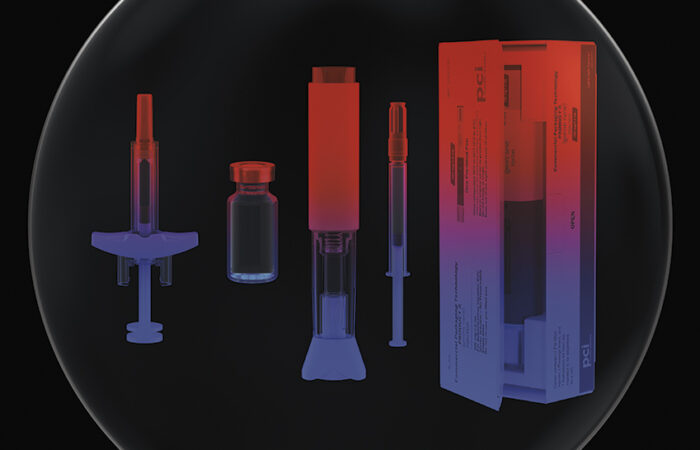
Gene therapy is delivered via one of two systems: viral or non-viral. These two methods can address all types of treatment purposes: suppression, enhancement or replacement of a disorder-related gene. Viral delivery systems are the most common delivery methods in US FDA-approved ocular gene therapy. A viral vector is delivered into or near the target tissues or cells, either by an injection, where the target is in the eye, or pharmaceutically, where the target is near the eye’s surface.
Viral vectors are built using a blueprint of a virus. They use the parts of the virus that help deliver genetic materials and exclude the parts that cause disease. They are widely considered to be more effective than non-viral vectors but do have some drawbacks – they are limited by their immunogenicity, oncogenicity and the small amount of DNA they can transport.
Adenoviruses, once a popular viral vector, have been largely abandoned for having strong immunogenicity and short expression duration. Lentiviruses bring an increased risk of oncogenesis (where healthy cells are transformed into cancer cells) because they naturally integrate into host genomes. Adeno-associated viruses (AAVs) are the most commonly used viral vector for ocular gene therapy. This is because they have three advantages over competing procedures:
- An ability to transduce multiple retinal cell types
- A relatively low immunogenicity
- Low oncogenicity because they do not integrate into the host genome like lentiviruses.
Non-viral vectors are less effective than viral vectors, but they are safer, less costly to develop, more reproducible and they do not have a DNA size limit issue. However, excluding those actively studied in vivo, non-viral delivery methods (either physical or chemical) have not been used as much as viral delivery systems.
“The purpose of ocular gene therapy treatment is to put genetic material into the target cells, so the delivery method depends on where those cells are located.”
The most actively researched non-viral vectors are chemical disruption, electroporation and polymer-based vectors. Electroporation is a physical form of non-viral delivery method and involves using pulses of electricity to create temporary pores in a cell membrane. These pores then enable genetic materials to be delivered into the cell to take effect. Electroporation has been explored in vivo, but the most recent applications have been on cells outside the body. The other method of delivering a non-viral vector is chemical, where lipid nanoparticles (LNPs) are used.
LNPs have membranes made from lipids similar to the molecules in cell membranes. Their main strength is versatility. They can be used to deliver DNA or mRNA, which can instruct cells to block a protein or make more of it. They have been described as a platform technology on which genetic solutions can be built quickly and loaded into the appropriate LNP.
When researchers are considering which kind of vector to use for gene therapy delivery, there are several things they must consider. Effectiveness, safety, the size of genetic materials, the duration of gene expression, the target cells, accessibility and cost all make the list of considerations. But the decision making does not end there. There is also the question of whether to use injected or pharmaceutical means to administer the ocular gene therapy. There are benefits and challenges to both but selecting the right one is crucial.
THE PHARMACEUTICAL APPROACH
Conventionally, drug delivery routes for treating an ocular disease have included systemic (oral delivery and intramuscular or intravenous injection), topical (eye drops, ointments) and injections in and around the eye.
The purpose of ocular gene therapy treatment is to put genetic material into the target cells, so the delivery method depends on where those cells are located. If they are near or on the eye’s surface, topical administration can work, but if the cells are in the eye, an injection is the best method.
Drug delivery via systemic or topical administration presents three major challenges. First, a tissue-specific delivery method needs to be developed. Secondly, doctors need to maintain a high local concentration of the genetic material. Lastly, the blood-retinal barrier can often block the drug from passing into the retina via systemic circulation.
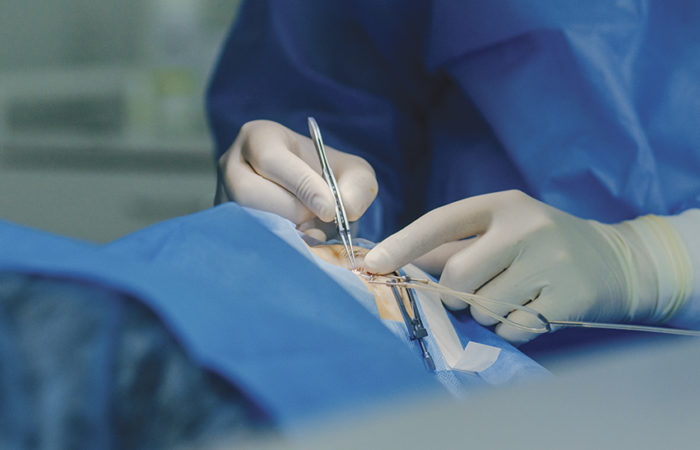
Most genetic diseases associated with the eye are either a form of retinopathy (a disease in the retina) or neuropathy (damage or disease in the nerves). The target cells in both cases are in the eye itself, so it is incredibly difficult to use a pharmaceutical approach, meaning that injections are necessary. However, injection is an invasive approach and comes with its own challenges.
THE INJECTION APPROACH
There are several different types of injections in and around the eye – subconjunctival, retrobulbar, intracameral, intravitreal, subretinal and suprachoroidal. Intravitreal and subretinal injections are the most popular administration routes, but others can be used, and new methods are emerging. For example, an intracameral injection can be applied if a genetic disorder involves the corneal endothelium, or if a gene therapy is being used for glaucoma.
The suprachoroidal injection route has been actively studied and potentially used in gene therapy for retinal or choroidal diseases, ocular oncology and steroid delivery. This new approach is being developed to target the posterior segment of the eye. This route has the potential to achieve chorioretinal concentrations 10 times greater than those obtained with intravitreal injections. It also avoids the need for vitrectomy and retinotomy, as is required for subretinal delivery. A suprachoroidal injection also reduces the drug’s exposure to the vitreous and anterior segment, potentially mitigating the side effect of increasing intraocular pressure found with steroids.
The process of administering an ocular injection is complex. A subretinal injection includes the following steps:
- First there is the vector preparation, then a transconjunctival puncture through the sclera and into the vitreous using a vitrectomy trocar.
- Next, leave the drive pipe in the sclera to facilitate the injection and conduct vitrectomy to remove the vitreous. If a vitrectomy is not conducted, the next step is a paracentesis of the anterior chamber, releasing aqueous humour for intraocular pressure control.
- After that, the practitioner must insert the 23-gauge subretinal injection needle into the vitreous, accessing the subretinal space while avoiding the major retinal vessels. At this point, the vectors are injected, generating a bleb beneath the neurosensory retina.
- Finally, the needle is pulled out, the drive pipe removed and the incision is sealed if necessary.
Challenges with Injections
Injections in such a delicate part of the body will always be complex. It can be even more challenging if the target cells are in the retina, which is a very subtle tissue. The procedure needs to be practiced often before it can be administered appropriately.
Scientists need to consider that while injection is currently the most common route for delivering gene therapies, it is not ideal. There is a risk of injury and infection from ocular injection. The most common injuries involve the iris, lens and retina and cause bleeding, cataract and retinal detachment. Intraocular pressure (IOP) needs to be monitored or prevented for intraocular injections by an anterior chamber paracentesis. A high IOP can cause damage to the retina and optic nerve, which is part of the mechanism of glaucoma.
To mitigate risks, trained surgeons or technologists should perform the procedure only after as much practice as possible. They should ensure that they are using qualified devices and instruments, conduct a strictly sterile operation to avoid infection and inflammation, monitor or prevent IOP, avoid injecting near blood vessels or touching the lens, and consider using steroids or immune-inhibiting medications.
If a non-invasive route exists, it is always preferable to something as invasive as an injection. Therefore, developing new non invasive means to deliver gene therapy would be highly worthwhile.
It is also important to consider how to best control toxicity when delivering ocular gene therapy. Producing too much toxicity can be mitigated by selecting the appropriate viral vectors, or non-viral vectors where suitable, a lower dose or concentration and applying an anti-inflammation treatment.
“Developing new non-invasive means to deliver gene therapy would be highly worthwhile.”
CONSIDERATIONS FOR DRUG DEVELOPERS AND SPONSORS
Drug developers and sponsors should consider all the above but also keep in mind a few other factors. They should note that the success rate of ocular injections and the level of experience with such procedures are critical when choosing a drug development partner.
On the practical side, they need to remember that spare animals may be needed for replacements in the case of failures, and steroids and immune-inhibiting medicines should be on hand to treat inflammation caused by the injection of vectors. And for viral vector delivery studies, a biosafety level-2 lab is required and may cost more when working with a lab testing partner.
It is also worth remembering that a number of factors determine the success of ocular gene therapy, some of which are out of the hands of drug developers. The effectiveness of gene therapy for ocular diseases can change depending on whether it is a single-mutation disorder or a multi mutation disorder. Then key decisions are made before treatment, which can also affect how a patient reacts. This can include establishing the appropriate method to ensure that the vectors are delivered to or near the target cells. It is also important to choose appropriate AAV capsids for gene therapy. Different serotypes of AAV provide different tropisms for different retinal cell types.
Other factors determining the outcome include the successful transduction rate of the vectors, the expression/transduction in the desired target cells and the expression duration. The patient’s immune reaction to the vectors will also determine success or failure, as will the proportion of functional cells remaining when the gene therapy is administered.
In vivo and in vitro studies suggest gene therapy will work best for people with single-mutation-induced disorders and more functional cells remaining and where there is high transduction, low immunogenicity and robust expression in the target cells.
A FINAL WORD ABOUT OCULAR GENE THERAPY
Ocular gene therapy is an exciting branch of medicine and promises to give hope to people worldwide suffering from conditions previously thought untreatable. The potential upsides for drug developers and sponsors interested in pursuing ocular disease treatments are substantial.
However, they must recognise the complexity of many factors, including delivery methods. Those looking into this topic seriously should first make a list of single-mutation disorders, explore orphan drug designation from the FDA as an option for rare genetic disease research, and choose a delivery system that ensures high transduction and low immunogenicity in the eye.
Finally, drug developers and sponsors need to ensure they collaborate with a team experienced in gene therapy studies and drug delivery techniques.