
Citation: Khatri A, Barbé CA, “Silica Nano-Solutions for Skin Delivery”. ONdrugDelivery Magazine, Issue 84 (Mar 2018), pp 12-16.
Aparajita Khatri and Christophe A Barbé outline the growth of the dermal delivery market, as well as the challenges it faces. The authors go on to detail a nanocomposite lidocaine patch, developed in collaboration with Nanopharma (Pardubice, Czech Republic), and how its achievements could be a boon to the skin delivery space as a whole.
CURRENT CHALLENGES IN SKIN DELIVERY
The development of dermal delivery systems has made pain-free self-administration a real option for patients. As such, patient acceptance of, and demand for, skin delivery products has increased, which have proven to be one of the main drivers for the skin delivery market. This market accounts for more than 12% of the overall global drug delivery market today.
Transdermal patches, ointments, gels and implants are currently the popular methods for transdermal drug delivery, with patches fast becoming the preferred option, especially for the elderly and children. Currently, 40 transdermal patches are available on the market, with over 70 more in clinical trials,1 pain relief and hormone therapy products being the most prevalent.
“While there has been considerable progress in the development of occluded and controlled-release pain relief systems, there remain some outstanding limitations, most notable of which is the undesirably high drug content during and after application…”
While there has been considerable progress in the development of occluded and controlled-release pain relief systems,2 there remain some outstanding limitations, most notable of which is the undesirably high drug content during and after application. In most cases, systems require a very high loading to obtain constant release of drug. Such high doses can trigger adverse skin reaction, thus limiting how long the system can be applied. This is highly inefficient as most of the drug remains in the patch after use (40-97%).
In addition to being uneconomical, inappropriate use and disposal of the patches also pose a serious public health risk, especially for controlled drug applications. In the case of fentanyl patches for example, although the clinically preferred treatment for several types of chronic pain, typically up to 60% of the drug is left in the patch when discarded. Inappropriate disposal, drug abuse and unintentional exposure are limiting the patches’ use in clinical medicine; lowered prescriptions have led to a drop of up to approximately 20% in annual sales of fentanyl patches. Furthermore, issues such as leakage of the drugs from the patch and/or crystallisation of the drug during application can lead to uncontrollable and unpredictable dose administration.
Another fast-growing facet of skin delivery based applications is bioactive wound management (BWM). It is forecast that the market for BWM will grow to US$4.9 billion (£3.5 billion) by 2020,3 a major driver for this being the use of growth factors. Growth factors help with wound regeneration and have the potential to reduce scarring.4 However, for these to be effective, sustained release is required during healing. This has been difficult to achieve thus far, due to these biomolecules being susceptible to rapid degradation by enzymes in the wound. The challenge of multiple high-dose administrations, frequent and painful dressing changes, and the high cost of treatment continue to impede the much needed commercialisation of these very promising bioactives.
SILICA PARTICLES FOR DRUG DELIVERY
Through a combination of sol-gel technology and emulsion chemistry,5 Ceramisphere can encapsulate a wide range of water soluble and insoluble actives inside an amorphous silica matrix. Biodegradable spherical matrix particles are produced under ambient conditions with a tailored size (from 50nm-100μm), providing controlled release of the active (from hours to days).6-8 The payload is released by diffusion through the 3D porous network of the silica “sponge”. The release rate is controlled by the internal structure of the sphere, which is customised during synthesis. Using the same base technology, the production processes have been scaled up to 100 kg/batch (for non-pharma products9) and are GMP compatible.
Ceramisphere has also developed specific processes to encapsulate a wide range of biological actives, e.g. small drug molecules (hydrophilic and lipophilic), polynucleotides (RNA/DNA) and therapeutic peptides and proteins.9 Efficient encapsulation is achieved (typically >80%) with no loss of structural integrity and function. The distinctive feature of this process is that the biomolecules are physically entrapped inside the silica matrix; this engenders excellent protection against degradative enzymes, acidic pH or temperature fluctuations. This encapsulation inside porous silica matrices is also known to prevent the crystallisation of poorly soluble drugs and enhance their bioavailability.10 In contrast to crystalline silica, amorphous silica has very low toxicity and is US FDA approved for non-injectable routes. In vitro dissolution studies using USP IV methodology have confirmed the biodegradability of Ceramisphere silica particles with dissolution ranging from hours (in vitro) to several days8 or weeks in vivo, depending on the route of administration and final target organ. The silica nanoparticles revealed an extremely low toxicity profile in mice, even at particle doses up to 200 mg/kg IV.9
In contrast to polymeric drug delivery technology, Ceramisphere silica particles are compatible with a wide range of post processing techniques. They are mechanically resistant and durable and are therefore able to survive grinding, milling, extrusion and electrospinning.
NANOCOMPOSITE SILICA-POLYMER PATCHES
Nanocomposite Patch Concept
Building on the key strengths of our silica delivery platform, such as protection and sustained release of the payload, and compatibility with spinning processes, Ceramisphere has developed the concept of a biodegradable nanocomposite patch in collaboration with Nanopharma AS. The nanocomposite patch is composed of biodegradable polymeric nanofibres containing silica nanoparticles with encapsulated bioactive(s). Produced by electrospinning, the production can be scaled up to meet industrial demand. The silica matrix provides both protection to the drug molecule during electrospinning and enables sustained release during application to the skin.
“Through a combination of sol-gel technology and emulsion chemistry, Ceramisphere can encapsulate a wide range of water soluble and insoluble actives inside an amorphous silica matrix…”
It is important to stress that silica encapsulation significantly enhances the functionality of these patches, offering the potential to introduce drugs which are incompatible with the polymeric chemistry or the spinning process into the nanofibrous system. These patches have a high safety profile, given that all components (polymer, silica matrix) are biodegradable, and FDA approved for skin applications.
Nanocomposite Lidocaine Patch
To demonstrate the potential of the nanocomposite patch and in particular its clinical potential, Ceramisphere and Nanopharma have developed a lidocaine nanocomposite patch. The choice of lidocaine was motivated by its excellent safety profile for the treatment of post-herpetic neuralgia (PHN) (Lidoderm®, Versatis®). In addition to potentially expediting the patch development to the clinical stage, this will also provide a clean baseline to showcase the potential of this delivery system.
Representing roughly a market of $1 billion (IMS Health, 2015), the lidocaine market continues to grow, driven mainly by an ageing population. With the European patent expiry in 2017, companies are actively looking for a differentiator technology that would give them an edge over generic competitors. The current lidocaine patch uses a very high drug load, with 97% remaining in the patch after use. It can only be applied for 12 hours with a subsequent 12 hour break, due to the high levels of lidocaine in the patch causing an adverse reaction in the skin.
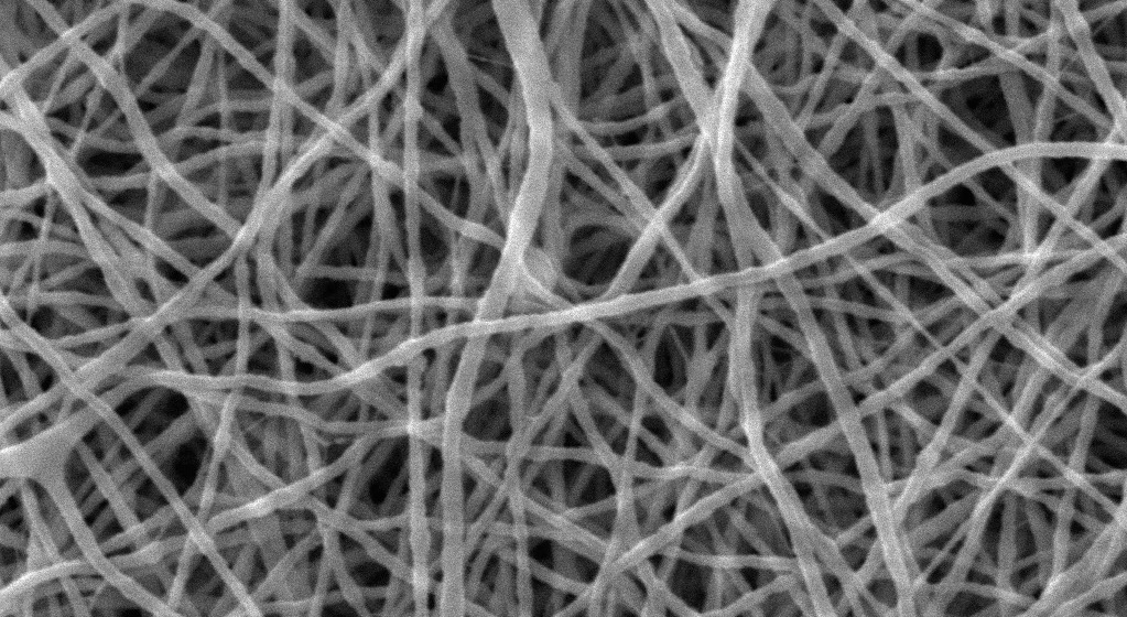
Ceramisphere and Nanopharma have produced a biocompatible nanocomposite lidocaine patch with the potential for a greater than 24 hour application and at least 30 times lower dose at the time of administration. The lidocaine is encapsulated inside the spherical particles (50-60 nm) of uniform morphology. The particles are then incorporated into the polymeric nanofibres using an electrospinning device to produce a non-woven nanocomposite mat (Figure 1).

Figure 1: Lidocaine silica particles (TEM) and the nanocomposite patch (SEM and Confocal). Confocal image shows fluorescent lidocane silica particles in nanofibres.
The features of the nanocomposite lidocaine patch are:
- Constant release: The encapsulation in silica matrix does not alter the release profile of lidocaine from the nanocomposite patch in vitro. When tested using a Franz diffusion cell (n=4), this release was found to be linear through the human skin and comparable to the commercial patch (Figure 2A).
- Faster pain relief: The Franz cell studies also showed a much shorter induction time of two hours between application and initial release, compared with over four hours for the commercial patch, suggesting faster pain relief. This was confirmed in animal studies, where the release of lidocaine in the blood plasma was evident as early as one hour after application of the nanocomposite patch versus over four hours for the commercial patch. Fast pain relief is ideal for paediatric analgesia, where transdermal patches are fast becoming the route of choice.
Figure 2: Comparison of patch performance in Franz cell studies using human skin.
- Superior efficiency: Defined here as the percentage of lidocaine released in comparison to the administered dose, this is one of the most attractive features of nanocomposite patches. In Franz cell studies, despite a between 60- and 100-fold lower lidocaine dose in the nanocomposite patch, approximately 80% of the administered lidocaine had passed through the skin after 24 hours (Figure 2B), which when combined with 17% present in the skin tissue (epidermis and dermis) (Figure 2C) suggested a total drug release of approximately 97% (i.e. less than 5% remaining in the patch after use). In comparison, a total of approximately 1% of lidocaine from the commercial patch was released. This was confirmed in large animal studies where, despite a 30-fold smaller dose in the nanocomposite patch, the delivery of lidocaine into blood plasma and the skin was comparable to the commercial patch (Figure 3).
- Low potential for skin toxicity: The nanocomposite patch is unlikely to lead to any specific nanoparticle based toxicity, as no evidence of skin penetration by fluorescent nanoparticles was found in vitro. This was further confirmed in animal trials, where no signs of any irritation/inflammation (redness) or swelling (Draize scoring) on the skin was recorded even after 72 hour application of the patches.
These nanocomposite patches are now ready for testing in humans and it is anticipated that clinical trials will prove these new patches to be safe and efficacious, providing a segue for new lidocaine products and posology.
Extension of the Technology to other Pain Relief Applications
In addition, this technology offers a clear solution to the issues dogging the delivery of opioid-based transdermal pain relief or other pain relief drugs. For example, a patch using silica with encapsulated fentanyl could be effectively administered using a much lower dose, with negligible quantities of drug remaining at the time of disposal. The danger of unintentional fentanyl release due to a temperature increase (e.g. fever) could potentially be minimised as silica-encased fentanyl will be protected against temperature fluctuations during application and storage of the patch.

Figure 3: Comparison of patch performance in piglets.
SILICA NANO-SOLUTIONS FOR WOUND MANAGEMENT
There is an opportunity to encapsulate growth factors, such as epidermal growth factor (EGF), in the silica matrix to protect them against wound mediated degradation (e.g. proteases) and control their release over time.
Ceramisphere has demonstrated that silica encapsulated EGF is functional, well protected in the wound environment and shows controlled release over a 14 day period (Figure 4A). In comparison to multiple doses of EGF, there is a clear advantage of the encapsulated EGF for re-epithelialisation of deep burn wounds at a faster rate, with potential for significant reduction in scarring (Figure 4B). This data underpins the current development of a silica particle-EGF nanocomposite patch and the development of silica particle-based smart wound dressings.

Figure 4: EGF silica particles for burn wound healing.
CONCLUSION
Silica particle-based delivery systems offer an overarching solution that tackles several of the challenges currently facing dermal delivery. Ceramisphere’s technology allows encapsulation and protection of a large range of bioactives and co-formulation of incompatible actives. This protection inside the silica matrix not only stabilises the drug, but also enhances its compatibility with other technologies, e.g. incorporation of other polymer fibres to produce a patch.
Capitalising on these features, Ceramisphere has developed a user-friendly and commercially viable skin delivery system with high functionality and high efficiency.
The nanocomposite patch technology can:
- Extend the time of application: Potentially over 24 hours for lidocaine patches, the skin irritation being minimised via drug encapsulation in the protective and biocompatible silica matrix.
- Increase the efficiency of the patch: More than 95% drug release demonstrated in the nanocomposite lidocaine patch, which could easily be extended to controlled drugs (e.g. fentanyl). The main features include significantly lowered dose loading and close to nil residual remaining in the used patch. This can also provide an economically viable and effective solution for expensive drugs (e.g. growth factors).
- Enable multidrug treatments in one device: Smart dressings with high functionalities can potentially be developed, allowing for multifaceted wound care, i.e. faster and better healing, low scarring and improved pain management. These systems are expected to have immediate application in skin grafts and for children, where there is a crucial need for pain-free wound healing solutions (e.g. for scalding and abrasions).
REFERENCES
- “Global Transdermal Patch Market & Clinical Pipeline Outlook 2022”. Kuick Research, March 2017. www.researchandmarkets.com/reports/4117865
- Pastore MN et al, “Transdermal patches: history, development and pharmacology”. Brit J Pharmacology, May 2015, 172, pp 2179-2209.
- “Bioactive Wound Care Market to Reach 4.9 Billion USD by 2020 – IndustryARC Research”. PR Newswire, Feb 2016. www.prnewswire.com/news-releases/bioactive-wound-care-market-to-reach-49-billion-usd-by-2020—industryarc-research-570105551.html
- Park JW, Hwang SR, Yoon IS, “Advanced Growth Factor Delivery Systems in Wound Management and Skin Regeneration”, Molecules, Jul 2017, Vol 22(8), epub.
- Barbé CJ, Bartlett JR, “Controlled release ceramic particles, compositions thereof, processes of preparation and methods of use”. International Patent Application WO2001/062232.
- Barbé CJ et al, “Sol–gel matrices for controlled release: from macro to nano using emulsion polymerisation”. J Sol-Gel Sci Technol, Jun 2008, Vol 46, pp 393-409.
- Finnie KS et al “Biodegradability of sol–gel silica microparticles for drug delivery”. J Sol-Gel Sci Technol, Jan 2009, Vol 49, pp 12-18.
- Finnie KS et al, “Encapsulation and controlled release of biomolecules from silica microparticles”. J Mater Chem, 2006, Vol 16, pp 4494-4498.
- “Silica for drug delivery”. Ceramisphere website. www.ceramisphere.com/healthcare/silica-for-drug-delivery/
- McCarthy CA et al, “Mesoporous silica formulation strategies for drug dissolution enhancement”. Expert Opin Drug Deliv, 2016, Vol 13(1), pp 93-108.
- Fleming JA, O’Connor BD, “Use of Lidocaine Patches for Neuropathic Pain in a Comprehensive Cancer Centre”. Pain Res Manag, Sep-Oct 2009, Vol 14(5), pp 381-388.
- Weil AB, Ko J, Inoue T, “The use of lidocaine patches”. Compend Contin Educ Vet, Apr 2007, Vol 29(4), pp 208-216. (NB Erratum in June 2007 Vol 29(6))