

Citation: Rupenthal ID, “Sector Overview Occular Drug Delivery Technologies: Exciting Times Ahead”. ONdrugDelivery Magazine, Issue 54 (Jan 2015), pp 7-11.
Ilva Rupenthal gives a sector overview of the occular drug delivery space.
SMALL ORGAN – BIG MARKET
The 2010 global cost of vision loss was nearly US$3 trillion (£2 trillion) for the 733 million people living with low vision and blindness,1 with this number expected to increase significantly due to the aging population and the increasing incidence of metabolic disorders such as diabetes. This makes eye disorders one of the costliest health conditions worldwide. The major blinding disorders currently include age-related macular degeneration (AMD), diabetic retinopathy / macular edema (DR/DME) and glaucoma, with these conditions offering great opportunities for innovation in drug delivery technologies. According to the Bright Focus Foundation, over 11 million people in the US have diagnosed AMD, with this number expected to double to nearly 22 million by 2050. Worldwide around 18 million injections of anti-VEGF agents were given in 2014 to treat AMD with an estimated global cost of visual impairment due to AMD being $343 billion, including $255 billion in direct health care costs. According to the International Diabetes Federation, around 387 million people are currently affected by diabetes with this number projected to increase by 205 million until 2035.
Within 20 years of diagnosis virtually all Type I diabetics and 60% of Type II diabetics show signs of retinopathy and according to the 2014 Prevent Blindness report, more than eight million people are currently affected by DR with this number projected to increase to nearly 11 million by 2032. Moreover, diabetes patients are 60% more likely to get cataracts and 40% more prone to develop glaucoma than those without diabetes, further increasing the incidence of ocular disorders in diabetics. Glaucoma currently affects over 2.8 million Americans with an estimated increase of 92% (a total of 5.5 million cases) expected by 2050. Today more than $6 billion is spent to treat glaucoma and optic neuropathies, with costs expected to double by 2032 and to rise to $17.3 billion by 2050 according to the 2014 Prevent Blindness report.
“The number of companies working on ocular drug delivery platforms, especially for the posterior segment, has skyrocketed over the past decade, with the reformulation of previously approved molecules offering the advantage of available long-term safety data in humans and therefore a facilitated approval process…”
While there are many effective medications to treat ocular conditions the challenge remains to deliver these drugs effectively with minimal side effects. Glaucoma eye drops, for example, usually need to be applied once or twice daily, often as a combination of multiple products, to achieve sufficiently high drug concentrations while intravitreal injections of antibodies against vascular endothelial growth factor (VEGF) in AMD treatment are generally given every 4-8 weeks. Therefore, achieving sufficiently high concentrations at the target site and maintaining these over prolonged periods with minimal side effects, offers great opportunities for new product development, especially when using already US FDA-approved drugs with well-known safety and efficacy.1
The global ophthalmology drug and device market was estimated at $36 billion in 2014 and is expected to increase to $52.4 billion by 2017. The pharmaceutical segment of this market was $19.8 billion last year, with AMD and DR drugs accounting for $5.84 billion.1 When comparing disease cases and revenue, anterior segment conditions (45% of cases) accounted for 95% ($4.9 billion) of the revenue in 2001. While the proportion of disease cases (45% anterior and 55% posterior segment) has not changed significantly since then, the revenue for posterior segment diseases is expected to increase from $0.3 billion in 2001 to about $9.9 billion by 2018 (reaching 43% of eye care costs) with the anterior segment accounting for the remaining 57% ($13.2 billion). Keeping these figures in mind it is no surprise that the number of companies working on ocular drug delivery platforms, especially for the posterior segment, has skyrocketed over the past decade, with the reformulation of previously approved molecules offering the advantage of available long-term safety data in humans and therefore a facilitated approval process, which will hopefully result in a number of innovative products on the market within the next few years.
SMALL ORGAN – BIG CHALLENGES
One would think that a small organ such as the eye which is readily accessible from the outside of the body would be easy to treat. However, the eye is a rather isolated organ with a number of barriers in place to protect it from the environment, which pose major challenges to effective drug delivery. When applying eye drops, for example, the most common method of treating anterior segment diseases, generally less than 5% of the applied drug reaches the ocular tissues. This is mainly due to the fast nasolacrimal drainage and the poor permeation of the remaining drug across the sandwich-like structure of the cornea, with the lipophilic corneal epithelium being the main barrier to ocular entry for most drugs. While this leads to excessive waste of costly drugs as well as low efficacy and patient compliance, it also poses a significant side effect risk due to systemic absorption of the majority of the dose given.
To overcome issues with topical ocular drug delivery researchers have focused predominantly on two strategies:
- Increasing the corneal residence time using viscosity enhancers, mucoadhesive, particulate and/or in situ gelling systems; and
- Increasing the corneal permeability using penetration enhancers, prodrugs and colloidal systems such as nanoparticles and liposomes.
While these have shown some improvements in the treatment of anterior segment diseases, they are unable to deliver sufficiently high concentrations to the back of the eye to treat most posterior segment conditions.
The gold-standard to treat conditions such as AMD is intravitreal injection of the drug-containing solution, although a few implants which are either sutured into the sclera or injected into the vitreous have also made it onto the market over the last two decades to treat a variety of retinal conditions (these will be further discussed below). However, although confined to the relatively small vitreal space, the drug faces elimination processes and needs to diffuse through the vitreous (with diffusion of large positively charged molecules hindered by the dense negatively charged vitreous meshwork) before crossing the inner limiting membrane to reach the retina with the RPE and Bruch’s membrane posing yet another permeation barrier for delivery into the choroid.
Other options to reach the choroid include periocular injections with the sclera allowing molecules up to 20 kDa to permeate (compared to ≤5 kDa across the cornea) as well as the possibility to inject larger volumes (≤1 ml compared with ≤100 μl intravitreally). Even more localised are suprachoroidal injections via precise microneedles which allow the drug solution to spread between the sclera and choroid around the eye ball, almost forming a drug-containing liquid ‘bandage’ around the eye. Again, there is a volume restriction (≤35 μl can be injected without leakage) and solutions are eliminated relatively quickly. However, injecting particles with a size smaller than 100 nm and/or in situ gelling systems into this space could offer sustained release possibilities.2
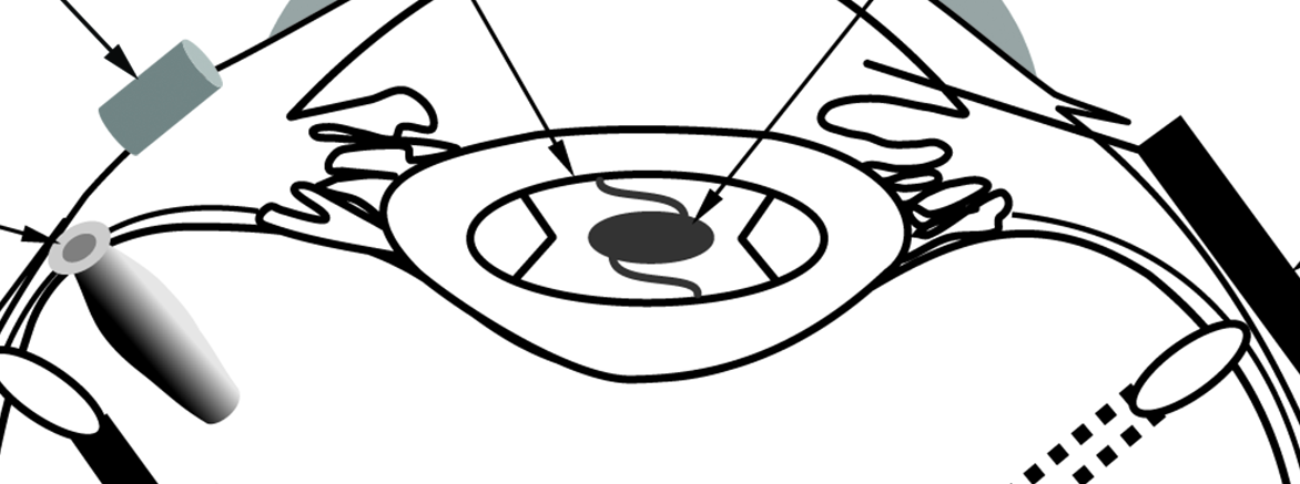
Finally, the active could also be administered systemically (oral or parenteral) and although choroidal blood flow is extremely high (up to 2000 μl/min/100 g of tissue with most other tissues having a rate of <500 μl/ min/100 g), generally less than 2% of the administered dose reaches the ocular tissues mainly owing to the tight blood-retinal barrier. However, a number of posterior segment diseases are characterised by leaky blood vessels which may result in higher drug concentrations in the effected ocular tissues and may increase the overall benefit to risk ratio after systemic administration, especially if the drug is encapsulated into a particulate system to protect it from degradation within the bloodstream. An overview of the ocular structures and recent delivery technologies, which will be further discussed below, is given in Figure 1.

Figure 1: Overview of the ocular structures and recent delivery technologies. (Modified with permission from Yasin et al.3)
RECENT ADVANCES IN ANTERIOR SEGMENT DELIVERY
Anterior segment diseases include blepharitis, conjunctivitis, corneal keratitis, dry eye, corneal infections and glaucoma with most of these currently treated with conventional eye drops such as solutions (ß-blockers, prostaglandin analogues (PGA), ß-agonists, carbonic anhydrase inhibitors and some antibiotics) or suspensions (steroids).
When taking anti-glaucoma medications as an example, only a limited number of advanced delivery systems have made it onto the market over the last decades. Allergan’s Propine® was the first prodrug-based eye drop to enter the market in the 1970s, with a 0.1% dipivalyl epinephrine solution lowering the intraocular pressure (IOP) as effectively as the 2% conventional solution. The closest to a microparticulate glaucoma formulation is Betoptic® S, which contains betaxolol hydrochloride bound to ion-exchange resin particles, with the 0.25% particle formulation found to be equivalent to a 0.5% Betoptic solution. A couple of in situ gelling systems are also available, including gellan gum based Timoptic® XE and xanthan gum based Timolol GFS®, both reducing the eyedrop application frequency from twice to once daily. Ocusert®, a pilocarpine-containing membrane-controlled reservoir system inserted into the conjunctival sac, offered sustained drug release over seven days. It was, however, relatively difficult to insert and often resulted in irritation and ejection.
In addition to these, a large number of exciting approaches are currently being researched or have entered into clinical trials. Novaliq GmbH, for example, is developing topical ocular formulations based on semi-fluorinated alkanes particularly suitable for the delivery of poorly soluble drugs such as Cyclosporin A (see this issue, Page 21). While enhancing drug dissolution and permeation and therefore increasing the overall drug bioavailability, the formulation does not require preservatives and lubricates the ocular surface, both aspects which are of particular benefit when treating dry-eye conditions. Kala Pharmaceuticals is developing mucus penetrating particles (MPP) which allow efficient penetration of the tear film mucin layer, the first defense mechanism encountered after topical administration of eye drops (see Issue 48, Page 16). This technology is currently under investigation to deliver loteprednol for a number of ocular conditions, with a twice-daily administration of the MPP-formulation as effective as four daily doses of the conventional suspension. Focusing again on novel antiglaucoma technologies, Envisia Therapeutics is investigating a PGA loaded PRINT-based biodegradable polymer rod intended for intracameral injection for three to eight months drug delivery (see Issue 48, Page 10), while GrayBug is developing proprietary PLGA particles (see this issue pp 24), which can release the IOP-lowering agent (GB-201) in a controlled fashion over several months, without causing any inflammation, a problem commonly associated with conventional particles.4 Currently in clinical trials are also three PGA-containing punctum plugs. The travoprost-containing plug from Ocular Therapeutix currently in Phase II clinical trials releases drug for up to 90 days and contains a visualisation aid to monitor plug retention. Both the QLT and Mati Therapeutics punctual plugs deliver latanoprost over three months and are currently in or have completed Phase II clinical trials.
“A recent study investigated nanodiamond-embedded contact lenses capable of lysozyme-triggered release of timolol maleate enabling drug release only once in contact with the tear fluid and therefore preventing premature drug release during storage…”
Drug-eluting contact lenses have also recently gained considerable interest for drug delivery to the anterior segment of the eye with the lens trapping the pre-corneal tear film and thus reducing its nasolacrimal drainage. Drug-loading approaches hereby include simple soaking of the lens in the drug solution (with limited sustained-release potential), incorporation of particles into soft contact lenses or molecular imprinting with the last two having shown drug delivery for up to one month. To reduce the initial burst and achieve controlled release, a recent study investigated nanodiamond-embedded contact lenses capable of lysozyme-triggered release of timolol maleate enabling drug release only once in contact with the tear fluid and therefore preventing premature drug release during storage.5
Besides refractive and drug delivery capabilities, soft contact lenses have also been investigated as sensors for glucose levels (Google) and intraocular pressure (Sensimed), but also have great potential to measure other clinical biomarkers in the tear film.6 The Triggerfish® from Sensimed AG, for example, is able to monitor the IOP telemetrically over 24 hrs.6 Similar to currently utilised glucose-sensing insulin pumps, one could therefore imagine a combination of sensing and drug eluting contact lens capabilities to achieve more efficient treatment of high pressure induced glaucoma. A similar concept is currently utilised in the Replenish MicroPump™ system, which contains a fluidic flow sensor, a bi-directional telemetry system for wireless programming and a microcontroller allowing pre-programmed administration of nanolitre-sized doses. With the addition of a pressure sensor, this technology could serve as a closed-loop system whereby an increase in the IOP would initiate on-demand drug release.7
RECENT ADVANCES IN POSTERIOR SEGMENT DELIVERY
Posterior segment diseases are the most prevalent cause of visual impairment in the developed world and include degenerative conditions such as AMD and retinitis pigmentosa, vascular diseases such as DR, retinal vein/artery occlusion and retinopathy of prematurity, inflammation such as seen in uveitis, infectious conditions including endophthalmitis and cytomegalovirus (CMV) retinitis as well as optic neuropathies arising from glaucoma.
The gold-standard to treat posterior segment conditions remains intravitreal injection which delivers the drug directly into the eye but may be associated with poor patient compliance and possible complications such a cataract formation and retinal detachment. Besides intravitreally injected solutions, four sustained-release implants have made it onto the market so far. These include non-biodegradable Vitrasert®, a ganciclovir containing scleral implant approved in 1996 for the treatment of CMV retinitis, and Retisert®, based on the same technology but smaller in size, which contains fluocinolone acetonide and was approved for the treatment of non-infectious posterior uveitis in 2005. Just recently US FDA-approved, Iluvien®, also a nonbiodegradable implant (polyimide tube with membrane caps), is injected into the vitreous with a 25G needle rather than being sutured into the sclera. It can deliver fluocinolone acetonide over three years for the treatment of DME, but little is currently known about its fate after drug depletion.
The first ever biodegradable intravitreal implant, Ozurdex®, was approved in 2009 and consists of a PLGA-based rod containing dexamethasone for the treatment of DME, retinal vein occlusion and posterior uveitis. The same technology is currently also under investigation for the delivery of brimonidine to treat dry AMD and retinitis pigmentosa, with the drug aimed at preventing cell death in the retinal pigment epithelium. It remains to be seen whether inflammation due to the low pH of PLGA degradation products, potentially masked when delivering an anti-inflammatory steroid (Ozurdex), may become more apparent with the brimonidine implant. GrayBug has therefore developed a proprietary PLGA–based technology proven to reduce the inflammation seen with conventional PLGA particles, with their lead product for wet AMD treatment (GB-102), exhibiting both anti-VEGF and anti-PDGF properties, targeted to last up to six months.4 The GrayBug technology allows relatively high drug loading and may also be capable of delivering large molecules including proteins, aptamers and biologics for several months.
In addition to Envisia’s intracameral implant discussed in the previous section, the PRINT technology has also been used for posterior segment systems. In the ENV705 implant a trehalose/bevacizumab mixture is dispersed within a polyglycolic acid matrix allowing drug release of effective concentrations over 3-6 months. While the Envisia particles are preformed, the Verisome® technology, based on carbonates, tocopherol and citrate esters, allows implant formation in situ once in contact with the aqueous vitreous environment. Depending on the volume of the formulation injected, drug levels can be maintained for up to 12 months with current clinical trials including dexamethasone and triamcinolone acetonide formulations.
“Whilst, to date, only open-loop systems responsive to light, or an electric or magnetic field have been explored for the purpose of drug delivery to the posterior segment of the eye pressure-responsive closed-loop systems may have great potential in the treatment of glaucoma and optic neuropathies…”
A rather different concept is utilised by Zordera which has developed a nanoporous film device with zero-order sustained drug release after intravitreal injection (see Issue 48, Page 20). This device consists of a drug pellet, sandwiched between two thin layers of impermeable biodegradable membrane, with adjustable nano-pores on one side permitting only one drug molecule to leave the reservoir at a time. The impermeable polymer layers degrade at a later time-point when most of the drug has been released, eliminating the need for device removal. This system has been shown to deliver ranibizumab over four months in a sustained manner.
While all of the above implantable systems may achieve sustained release, the release rate can generally not be altered if the condition worsens or aborted in the case of serious side effects. A refillable (to increase the device’s life span) and flushable (to abort drug delivery) functionality has, however, been included in the port delivery system, a scleral plug consisting of a porous container, a semi-permeable non-biodegradable membrane with a refill port and one or more exit ports to release the drug into the vitreous humour. This implant has been tested with ranibizumab and exhibited significant positive clinical outcomes when compared with monthly intravitreal injections of the same drug. With advances in polymer science and nanomaterial development for biomedical applications over recent years there has been a paradigm shift from conventional to stimuli-responsive or tunable devices, with such systems also having great potential in the area of ocular drug delivery.3
Whilst, to date, only open-loop systems responsive to light (e.g. ODTx), or an electric or magnetic field (e.g. micro-electro mechanical systems (MEMS)), have been explored for the purpose of drug delivery to the posterior segment of the eye,3 pressure-responsive closed-loop systems may have great potential in the treatment of glaucoma and optic neuropathies. Similar to the anterior MicroPump discussed in the previous section, the posterior Replenish system may be suitable to deliver neuroprotective agents to the posterior segment to prevent retinal ganglion cell loss resulting from elevated IOP. In the future, other sensing capabilities such as the measurement of VEGF levels, inflammatory markers or fluid build-up in the macula combined with a feedback mechanism and stimuli-responsive drug release could offer tailored and efficient treatment of posterior segment diseases.
CONCLUSION
A number of innovative drug delivery technologies are currently in the pipeline to treat ocular diseases more efficiently while minimising side effects, with the majority aiming to sustain drug release over prolonged periods. This would reduce the overall administration frequency and the need for specialist visits in the case of intravitreal injections, significantly reducing treatment costs and improving the quality of life for those affected by vision-impairing and blinding diseases. However, long-term effects of low sustained drug levels especially in the posterior segment are currently unknown and tunable drug release may be of advantage if the condition improves to avoid side effects such as the increased incidence of geographic atrophy seen when comparing monthly to as needed intravitreal injections of ranibizumab over 24 months.8 Thus, while sustained release is certainly of great advantage, variations in the disease progression, individual patient response to treatment and concurrent conditions may require alterations in the drug release pattern.
Tunable systems with the addition of a sensing capability may therefore offer more effective treatment of ocular diseases in the future. The major hurdle with successful development of such tunable implants remains the device size restriction, which in turn limits the amount of drug loading. Thus, to achieve sustained release over prolonged periods, such devices are generally limited to highly potent drugs such as steroids and may become difficult to adapt to macromolecules such as anti-VEGF agents. Moreover, matching these new technologies with drug pharmacokinetics and translating pre-clinical studies into human clinical trials remains a challenge. Nevertheless, novel ocular drug delivery technologies offer great market potential and it is certainly a very exciting area to be working in at the present time.
REFERENCES
- O’Rourke M, “Next Generation Ocular Drug Delivery Platforms”. Eye on Innovation, The Official Publication of OIS, 2014, Vol 1, p 7.
- Albini TA, Moshfeghi AA, Yeh S, “A Peek down the pipeline: Emerging drug delivery options for retinal disease”. Retina Today July/August 2014, pp 70-77.
- Yasin MN, Svirskis D, Seyfoddin A, Rupenthal ID, “Implants for drug delivery to the posterior segment of the eye: A focus on stimuli-responsive and tunable release systems”. Journal of Controlled Release, 2014, Vol 196, pp 208-221.
- O’Rourke M, Hanes J, “Ocular Drug Delivery Via Micro- and Nanoparticles”. Retina Today, 2014, July/August, pp 84-87.
- Kim HJ, Zhang K, Moore L, Ho D, “Diamond Nanogel-Embedded Contact Lenses Mediate Lysozyme- Dependent Therapeutic Release”. ACS Nano, 2014, Vol 8(3), pp 2998-3005.
- Farandos NM, Yetisen AK, Monteiro MJ, Lowe CR, Yun SH, “Contact Lens Sensors in Ocular Diagnostics”. Advanced Healthcare Materials, 2014 (DOI 10.1002/adhm.201400504).
- Boyer DS, Campochiaro PA, Humayun MS, Schopf L, Vold SD, Wirostko BM, “Emerging Technologies Approach Complex Eye Diseases from New Directions”. Ocular Surgery News APAO, 2014, May, pp 8-22.
- Martin DF, Maguire MG, Fine SL, Ying G-s, Jaffe GJ, Grunwald JE, Toth C, Redford M, Ferris 3rd FL, “Ranibizumab and Bevacizumab for Treatment of Neovascular Age-related Macular Degeneration: Two-Year Results”. Ophthalmology, 2014, Vol 119(7), pp 1388-1398.